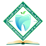
About the Laboratory:
The Radiology Laboratory at the College of Dentistry is a specialized facility dedicated to training students in radiographic and facial imaging techniques. It supports the diagnostic process in oral and dental surgery according to the latest scientific and professional standards. The laboratory operates within the framework of quality assurance and academic accreditation, adhering to Standard Operating Procedures (SOPs) and strict radiation safety and protection instructions.
Definition of Dental Radiology:
Dental radiology involves the diagnostic use of X-rays to detect: Interproximal caries • Periapical infections • Alveolar bone loss • Impacted molars • Orthodontic and surgical treatment planning.
Laboratory Objectives:
The lab aims to provide high-efficiency practical training on dental X-ray units, develop skills in interpreting radiographic images for pathological diagnosis, and reinforce radiation protection principles for both patients and operators. Furthermore, it supports research activities in the field of dental radiographic imaging.
Core Goals of the Radiology Unit:
1. Practical student training and interpreting radiographic images.
2. Implementing radiation protection principles and ensuring image quality with the minimum possible dose.
3. Qualifying students for clinical practice according to international standards.
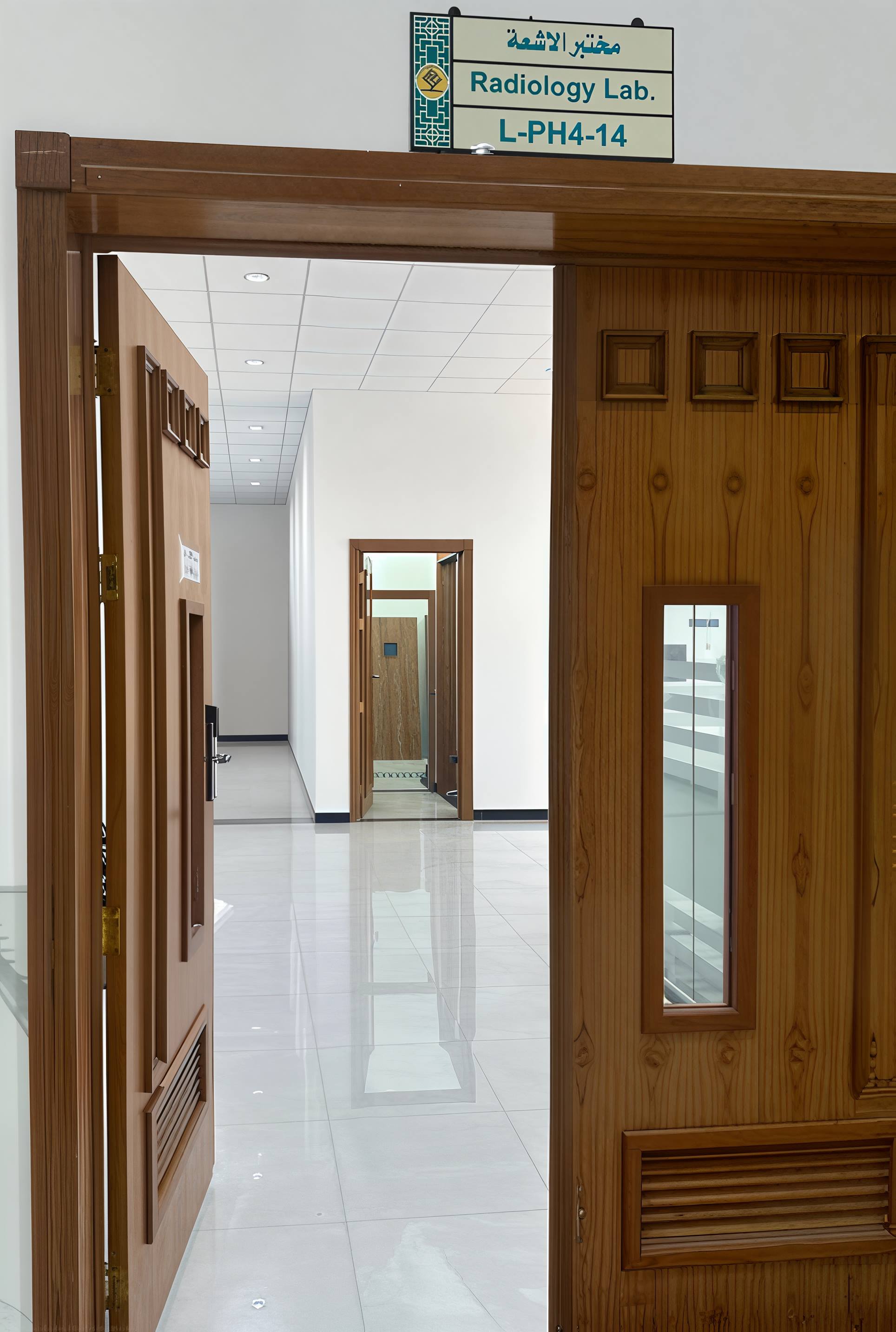
Diagnostic Radiographic Imaging:
• Periapical X-ray: Single dental images to evaluate teeth and roots.
• Panoramic X-ray: Full jaw imaging to identify anomalies, fractures, and bone diseases.
• Cephalometric X-ray: Lateral skull images for bone growth studies and orthodontic planning.
• CBCT: High-resolution 3D Cone Beam Computed Tomography for advanced diagnostic needs.
Radiation Safety Standards:
Commitment to safety guidelines involves using personal protective equipment (PPE) such as lead aprons and exposure-limiting devices, while maintaining a secure work environment through rigorous student training on preventive procedures.
Primary Functions and Capacity:
The unit provides organized training under specialized supervision, periodic equipment maintenance, and quality control documentation. The educational capacity is optimized at 10–15 students per session, divided into small groups to ensure direct supervision and prevent overcrowding in the X-ray room for safety purposes.
Academic Quality and Safety Measures:
This includes periodic equipment calibration, recording radiation exposure doses, training on emergency protocols, ensuring patient data confidentiality, and implementing a comprehensive Quality Control Program.
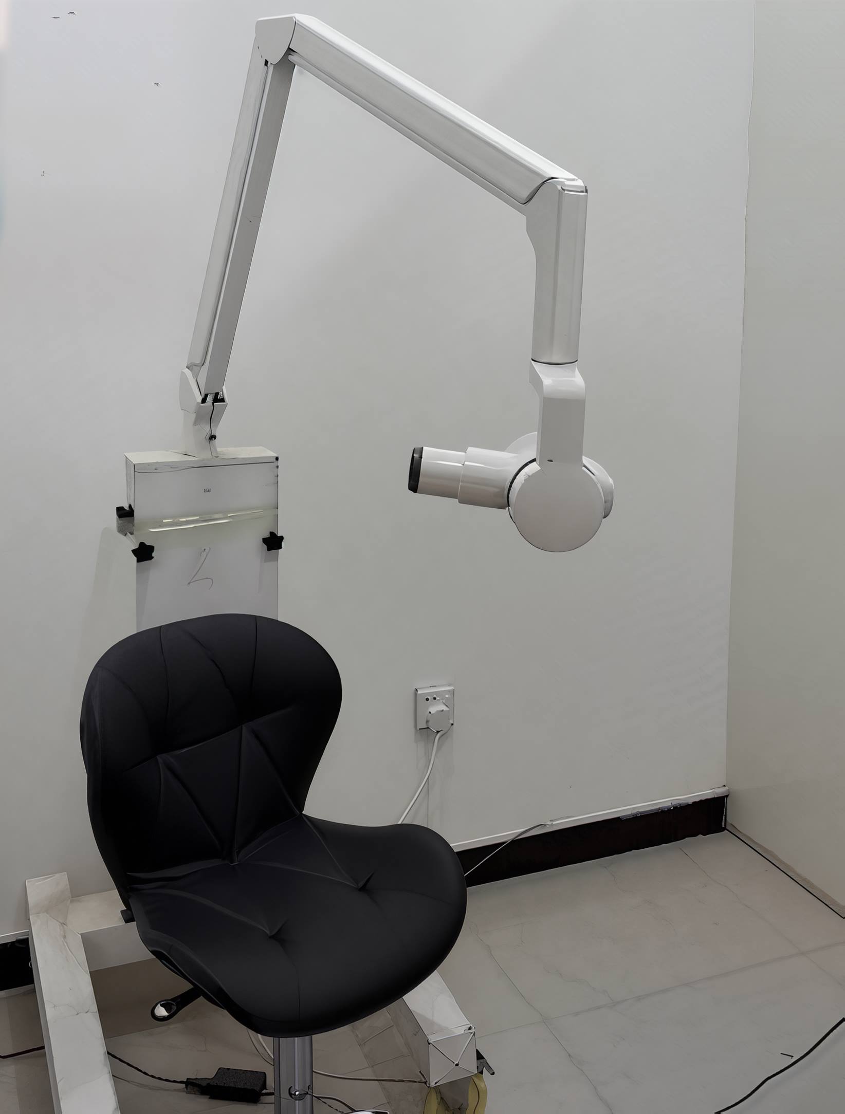
Essential Safety and Educational Equipment:
• Lead Apron and Thyroid Shield • Fixed Lead Barriers • Dosimeters • Radiation Warning Signs • Dental X-Ray Units • OPG and CBCT Units • Digital Sensors • Imaging Software.